Oncologic Imaging: PET-CT, MRI, and Staging Accuracy Explained
 Jan, 23 2026
When cancer is suspected, doctors don’t just guess where it is or how far it’s spread. They rely on imaging to see the invisible. Three tools dominate this process: PET-CT, MRI, and the newer hybrid PET-MRI. Each gives different information, and choosing the right one can change everything - from treatment plans to survival odds.
What PET-CT Actually Shows
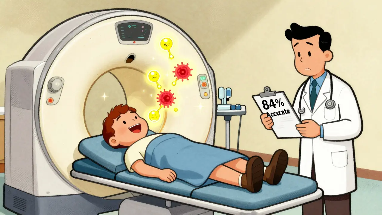
PET-CT is the most common tool for cancer staging. It combines two scans into one: a PET scan that shows where cells are using sugar at high rates (a sign of cancer), and a CT scan that maps out the body’s anatomy. Together, they paint a picture of not just where a tumor is, but how active it is.
The tracer used is usually 18F-FDG - a radioactive sugar. Cancer cells gobble it up faster than normal cells, lighting up on the scan. A typical adult dose is 370 MBq. The CT part gives detail down to half a millimeter, so doctors can pinpoint the tumor’s exact size and location.
For lung cancer, lymphoma, and melanoma, PET-CT is often the first choice. A 2023 meta-analysis found it correctly identified cancer spread in lymph nodes for non-small cell lung cancer with 84% accuracy. It’s fast - most scans take 15 to 20 minutes. And because it’s widely available, it’s the default in most hospitals.
But it’s not perfect. PET-CT can’t always tell the difference between cancer and inflammation. A swollen lymph node from an infection might light up just like a tumor. Radiation exposure is another concern: a full-body PET-CT gives 10 to 25 mSv - roughly 3 to 8 years’ worth of natural background radiation. That’s why it’s not used lightly in younger patients.
MRI: The Soft Tissue Champion
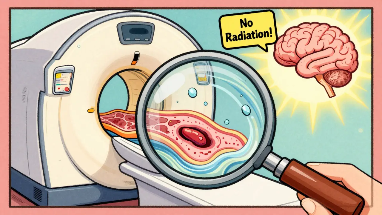
If PET-CT shows you where the cancer is, MRI tells you what it’s doing to surrounding tissue. No radiation. No tracers. Just powerful magnets and radio waves that reveal subtle differences in water content and cell structure.
At 3 Tesla, modern MRI machines can resolve details as small as 0.5 mm. That’s critical for cancers hidden in soft tissue - like prostate, liver, brain, or pelvic tumors. For prostate cancer, multiparametric MRI detects tumors with 75% accuracy, outperforming PSMA PET-CT in some studies. In breast cancer, MRI is the most sensitive tool for spotting hidden tumors, especially in dense tissue.
Functional MRI techniques like diffusion-weighted imaging (DWI) add another layer. They measure how water moves inside tissues. Cancer cells are packed tightly, so water doesn’t flow freely - that’s visible on the scan. This helps distinguish tumors from scars or fluid buildup after treatment.
But MRI has downsides. It takes longer - 30 to 60 minutes. Patients have to lie perfectly still. Motion ruins the images. People with pacemakers, metal implants, or severe claustrophobia often can’t have one. And while it’s great for anatomy, MRI doesn’t show metabolic activity like PET does. That’s why it’s often paired with PET.
PET-MRI: The Hybrid Advantage
PET-MRI came onto the scene in 2011, combining the metabolic power of PET with the soft-tissue clarity of MRI. It’s not just a mashup - it’s a new kind of diagnostic tool.
Because both scans happen at the same time, there’s no misalignment between the metabolic and anatomical images. That’s huge for brain tumors. Radiation necrosis (tissue damage from treatment) looks a lot like tumor recurrence on MRI alone - but PET-MRI can tell them apart with 85-90% accuracy, compared to 70-80% for MRI by itself.
For liver metastases, pelvic cancers, and pediatric tumors, PET-MRI is becoming the gold standard. A 2023 study in RadioGraphics found that PET-MRI changed treatment plans for nearly half of pancreatic cancer patients because it spotted small tumors MRI missed and ruled out false positives PET-CT flagged.
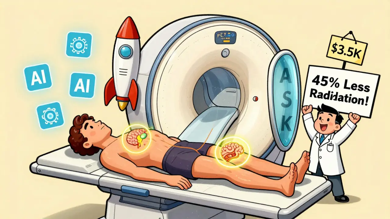
It also cuts radiation exposure by about half. That’s a big deal for kids, young adults, and anyone needing repeated scans over years. A child with neuroblastoma might get 10 PET scans over a decade. Switching to PET-MRI reduces their lifetime radiation dose dramatically.
But PET-MRI isn’t for everyone. It’s expensive - $2,500 to $3,500 per scan in the U.S., compared to $1,600-$2,300 for PET-CT. The machines cost $3-4.2 million to install. Only about 1 in 5 U.S. cancer centers have one. And the scan takes longer - 45 to 60 minutes. Motion artifacts are common. Technologists need extra training. Many hospitals still struggle with reimbursement.
Which One Should You Get?
There’s no one-size-fits-all answer. The choice depends on the cancer type, the question being asked, and what’s available.
- For lung, lymphoma, or melanoma staging: PET-CT is still the go-to. Fast, proven, widely available.
- For prostate, liver, or pelvic cancers: MRI or PET-MRI. Better at spotting small tumors and distinguishing cancer from scar tissue.
- For brain tumors or suspected recurrence after radiation: PET-MRI wins. It’s the only way to reliably tell if it’s cancer coming back or just radiation damage.
- For children or young adults needing long-term monitoring: PET-MRI is preferred to avoid unnecessary radiation.
- For emergency or frail patients: PET-CT’s speed and availability make it the practical choice.
Experts like Dr. Richard L. Wahl from Johns Hopkins say PET-MRI isn’t replacing PET-CT - it’s complementing it. Think of it like this: PET-CT is your flashlight in the dark. MRI is your magnifying glass. PET-MRI is both, in one hand.
What’s Changing in 2026?
The field is moving fast. In January 2024, Siemens got FDA clearance for a new PET-MRI system that cuts scan time to just 6 minutes for a whole-body scan. That’s a game-changer for patients who can’t lie still for an hour.
New tracers are also making waves. PSMA-targeted PET tracers (like 68Ga-PSMA-11) are now standard for prostate cancer. They light up cancer cells with far greater precision than old FDG tracers. When combined with multiparametric MRI, they’re changing how we stage and treat prostate cancer.
Artificial intelligence is stepping in too. At the 2023 RSNA meeting, researchers showed AI models that predict how a tumor will respond to chemo - based only on PET-MRI images. In trials, these models outperformed human radiologists in spotting early signs of treatment failure.
Guidelines are catching up. The 2024 ASCO recommendations now tell doctors to choose imaging based on tumor biology - not just location. For example, triple-negative breast cancer responds better to PET-CT early on, while hormone-receptor-positive tumors show clearer changes on MRI.
Real-World Challenges
Even the best technology doesn’t help if it’s not used right.
A 2022 survey of 127 radiologists found that 68% trusted PET-MRI more for liver lesions - but 73% said the long scan time made scheduling impossible. One tech on Reddit said their PET-MRI machine sits idle 40% of the time because staff aren’t trained to run it efficiently.
Attenuation correction - the process of adjusting PET data for tissue density - is a headache in PET-MRI. Without accurate CT data, the PET images can be wrong. Sites using advanced algorithms like Dixon-based correction report fewer errors, but it requires physics support most small hospitals don’t have.
Cost is the biggest barrier. In the U.S., Medicare and private insurers still reimburse PET-MRI at the same rate as PET-CT - even though the procedure costs 50% more. Many centers lose money on every PET-MRI scan. That’s why it’s mostly found in academic hospitals, not community clinics.
Still, adoption is growing. The global PET-MRI market is projected to hit $1.1 billion by 2030. More training programs are popping up. The EANM and ESMRMB now recommend PET-MRI for specific cancers. And as AI automates analysis and scan times shrink, the barriers will fall.
What Patients Should Know
If you’re facing cancer imaging, ask: What question are we trying to answer?
Are we looking for spread? Then PET-CT might be enough.
Are we trying to see if a liver spot is cancer or just a benign cyst? Then MRI or PET-MRI is better.
Are you young? Or do you need scans every few months? Ask if PET-MRI is an option to reduce radiation.
Don’t assume the most advanced test is always best. The right test is the one that answers your doctor’s question - clearly, safely, and quickly.
Husain Atther
January 24, 2026 AT 14:21PET-CT has been a game changer in my clinic, especially for lymphoma cases. The speed and clarity make it ideal for initial staging, even if we sometimes have to follow up with MRI when inflammation is suspected. It's not perfect, but it's reliable and fast - and in oncology, time matters.
Still, I'm glad we're seeing more PET-MRI adoption. The reduced radiation for younger patients is a big win, and the soft tissue contrast is unmatched. We just need better reimbursement and training to make it scalable.
Izzy Hadala
January 26, 2026 AT 05:36The integration of PSMA-targeted tracers with multiparametric MRI represents a paradigm shift in prostate cancer diagnostics. The sensitivity and specificity metrics reported in recent literature are statistically compelling, particularly when compared to conventional FDG-PET/CT. Moreover, the convergence of quantitative imaging biomarkers with AI-driven predictive modeling suggests a future where imaging not only detects disease but anticipates therapeutic response.
It is imperative that clinical guidelines evolve in tandem with technological advancements to ensure equitable access and evidence-based application across healthcare systems.
Marlon Mentolaroc
January 27, 2026 AT 02:25Let’s be real - PET-CT is the bread and butter because it’s cheap and fast. But the real magic? PET-MRI. I’ve seen cases where PET-CT said ‘probably metastasis’ and PET-MRI said ‘nah, that’s just scar tissue from last year’s surgery.’ Saved a guy from unnecessary chemo.
Now the problem? Hospitals charge the same for both, so they’d rather keep the old machine running and ignore the $4M shiny new toy in the corner. It’s not about tech - it’s about money. Always money.
Gina Beard
January 28, 2026 AT 20:10Imaging is not just seeing. It’s interpreting silence.
The body speaks in water diffusion, in glucose uptake, in magnetic resonance. The machine listens. The radiologist translates. And the patient? They just want to know if it’s gone.
Perhaps the real question isn’t which scan to use - but whether we’re listening closely enough.
Don Foster
January 29, 2026 AT 22:01Anyone who still thinks PET-CT is the gold standard hasn't read the 2023 RadioGraphics paper on pancreatic cancer. PET-MRI changes everything. The attenuation correction issue? Solved with Dixon algorithms. The cost? Worth it when you avoid a false positive that leads to a laparotomy. And don't even get me started on how FDG PET misses low-grade tumors. PSMA is the future and anyone who disagrees is stuck in 2015
Also AI is already outperforming radiologists in detecting chemo response. Wake up.
siva lingam
January 30, 2026 AT 08:44So we got a 4 million dollar machine that takes an hour, costs twice as much, and sits unused 40% of the time... because we're too lazy to train staff
Meanwhile the guy with lung cancer still gets the 15 minute PET-CT because someone actually knows how to run it
Progress is just expensive bureaucracy with better lighting
Shelby Marcel
January 30, 2026 AT 15:38wait so pet-mri is better but like... no one uses it because its expensive and slow?? and they still pay the same?? lmao
so we're basically paying for a fancy car but only using the radio
also i think i saw a post on r/radiology where someone said the techs call it 'the patience tester' 😅
blackbelt security
February 1, 2026 AT 13:25Every scan is a step toward hope. Even if it’s just a shadow on a screen, it’s a clue. A chance. A path.
Don’t let cost or convenience silence the potential of better tools. The future isn’t in the machines - it’s in the choices we make to use them.
Keep pushing. Keep learning. Keep asking for more.
Josh McEvoy
February 3, 2026 AT 09:48pet-mri = 🚀
but also = 💸💤
and also = 🤯 when it works
my uncle got scanned with it last year - turned out the 'tumor' was just inflammation from a weird infection
saved him from chemo, surgery, and a 3 month spiral into depression
so yeah... it's worth it. even if the machine takes a nap between patients 😴